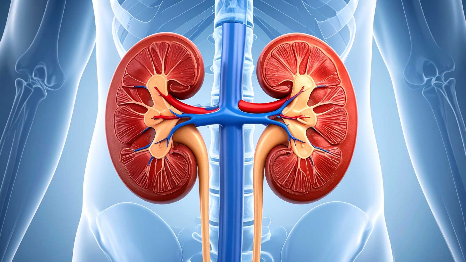
Pediatric Kidney Disorders: Early Detection and Modern Treatment
Introduction
Globally, congenital anomalies are among the major causes of childhood illness. According to the World Health Organization (WHO), congenital conditions account for a significant proportion of neonatal morbidity. Early screening and modern surgical technologies are therefore crucial for improving child health outcomes.
Global and India Context: Congenital Anomalies
| Indicator | Global Situation | India Situation | Source |
|---|---|---|---|
| Newborn deaths due to congenital disorders | ~240,000 deaths annually within the first 28 days | Data varies by condition; congenital anomalies are among the leading causes of neonatal mortality | WHO |
| Share of kidney & urinary tract anomalies | ~20–30% of prenatal abnormalities detected in ultrasound | Similar trend observed in prenatal screenings in Indian hospitals | Medical studies |
| Annual births | ~130 million births globally each year | ~25 million births annually | UN / Government of India |
| Children born with birth defects | Millions globally affected each year | ~1.7 million children affected annually | March of Dimes |
India carries a large share of the global burden of birth defects due to its high birth rate, making early screening and pediatric healthcare systems critical for child health policy.
“Prevention is better than cure.” — Desiderius Erasmus
This principle underpins modern approaches to prenatal screening and pediatric healthcare.
Importance of Antenatal Screening
Antenatal ultrasound has become an essential tool in maternal and child healthcare.
| Aspect | Significance |
|---|---|
| Early detection | Identifies congenital abnormalities before birth |
| Monitoring | Enables continuous tracking of fetal development |
| Timely treatment | Allows early medical or surgical intervention |
| Parental counselling | Helps families prepare for possible treatment |
In many cases, abnormalities detected during pregnancy may resolve naturally as the child develops.
Hydronephrosis: Most Common Prenatal Finding
One of the most commonly detected kidney conditions in prenatal scans is hydronephrosis, which refers to dilation of the renal pelvis.
| Feature | Details |
|---|---|
| Nature | Enlargement of the kidney’s urine-collecting region |
| Detection | Antenatal ultrasound |
| Outcome | Often resolves naturally after birth |
| Management | Monitoring through follow-up scans |
Doctors frequently adopt a “watchful waiting” approach, since many cases improve without medical intervention.
Posterior Urethral Valves (PUV)
Some congenital conditions may require early medical intervention.
| Aspect | Explanation |
|---|---|
| Condition | Posterior Urethral Valves |
| Occurrence | Only in male infants |
| Cause | Obstructive tissue in urethra |
| Impact | Urine backup causing pressure on kidneys |
| Treatment | Endoscopic removal using cystoscopy |
Modern pediatric urology uses minimally invasive endoscopic techniques, reducing recovery time and complications.
Vesicoureteral Reflux (VUR)
Recurrent urinary infections in children may indicate vesicoureteral reflux, where urine flows backward toward the kidneys.
| Feature | Explanation |
|---|---|
| Cause | Weak valve between ureter and bladder |
| Symptom | Recurrent urinary tract infections |
| Risk | Kidney damage if untreated |
Treatment Methods
| Method | Description |
|---|---|
| Endoscopic injection | Gel placed to prevent urine backflow |
| Laparoscopic surgery | Corrects position of ureter |
These techniques ensure short hospital stays and faster recovery.
Pelviureteric Junction (PUJ) Obstruction
In older children, a blockage between the kidney and ureter may cause pain.
| Feature | Description |
|---|---|
| Age group | Often seen in children aged 5–15 years |
| Symptom | Pain in the side or back |
| Treatment | Surgical correction |
Role of Advanced Surgical Technology
Modern pediatric surgery increasingly uses robot-assisted procedures, offering several advantages.
| Benefit | Explanation |
|---|---|
| Precision | 3D magnified surgical view |
| Minimal incisions | Reduced scarring |
| Faster recovery | Shorter hospital stays |
These innovations represent a major shift toward child-friendly and minimally invasive healthcare.
Comparison: Traditional vs Modern Pediatric Surgery
| Parameter | Traditional Surgery | Modern Techniques |
|---|---|---|
| Incision size | Large cuts | Small keyhole incisions |
| Recovery time | Longer | Faster |
| Hospital stay | Extended | Short |
| Surgical precision | Limited | High |
Medical technology has thus improved treatment outcomes significantly.
Relevance for Public Health Policy
Early diagnosis and treatment of congenital kidney disorders contribute to broader health goals.
| Public Health Impact | Outcome |
|---|---|
| Reduced kidney damage | Improved child health |
| Lower infection rates | Better quality of life |
| Early intervention | Prevention of chronic kidney disease |
India’s healthcare policies emphasise maternal and child health screening programmes, which play a vital role in detecting congenital conditions early.
Policy Relevance in India
India has introduced several programmes aimed at improving child health.
| Programme | Objective |
|---|---|
| Rashtriya Bal Swasthya Karyakram (RBSK) | Early detection of birth defects and childhood diseases |
| National Health Mission | Strengthening maternal and child healthcare |
| Ayushman Bharat | Expanding access to affordable treatment |
These initiatives support early screening and specialised pediatric care.
Conclusion
Advances in prenatal screening and minimally invasive surgery have transformed the management of congenital kidney disorders in children. Early diagnosis allows doctors to monitor and treat conditions before they become severe, significantly improving long-term health outcomes.
Strengthening antenatal care, expanding access to pediatric surgical technology, and improving public health programmes will be critical in ensuring healthier childhood development.
“The greatest wealth is health.” — Virgil
Attribution
Original content sources and authors
Syllabus classification
How this article maps to GS papers
Main syllabus
GS2HealthcareQuick Q&A
What is prenatal kidney anomaly detection, and how has modern medical technology transformed the diagnosis of kidney-related conditions in unborn babies?
The importance of this technological advancement lies in the shift from reactive to preventive and planned healthcare. Once a possible anomaly is detected, doctors can closely monitor the development of the foetus through follow-up scans and provide counselling to parents. In many cases, what initially appears as an anomaly is merely a temporary developmental variation that resolves naturally after birth. Thus, early diagnosis often prevents unnecessary panic and enables doctors to prepare a tailored care plan if intervention becomes necessary.
Example: A common condition detected through prenatal ultrasound is hydronephrosis, where the renal pelvis appears dilated. In most cases, doctors simply monitor the baby after birth with follow-up scans. This illustrates how modern diagnostics help transform a potentially alarming discovery into a manageable clinical observation. Ultimately, prenatal detection represents a major leap in paediatric healthcare by ensuring that infants receive timely and targeted care from the very beginning of life.
Why is early diagnosis of kidney-related conditions during pregnancy considered beneficial for both doctors and parents?
From the perspective of parents, early diagnosis helps reduce uncertainty and enables informed decision-making. Although hearing about a potential anomaly can initially create anxiety, it also provides reassurance because doctors can track the condition and explain possible outcomes. In many cases, the detected abnormality is mild and resolves naturally as the baby grows. The ability to conduct follow-up scans after birth ensures that any developing issue can be identified and addressed early, thereby preventing long-term complications such as kidney damage or recurrent infections.
Real-world implication: Suppose a prenatal scan detects mild dilation in the baby’s kidney. Instead of waiting until the child develops symptoms like fever or urinary infection, doctors can monitor the condition through postnatal ultrasounds. If the dilation reduces naturally, no intervention is required. If it persists, timely treatment can be provided. Therefore, early diagnosis serves as both a reassurance mechanism and a preventive healthcare strategy, significantly improving outcomes for newborns.
How does hydronephrosis develop in unborn babies, and what is the typical medical approach to managing this condition?
The standard medical approach in most cases is known as “watchful waiting.” Rather than performing immediate surgery or intervention, doctors carefully monitor the baby’s condition through follow-up ultrasound scans after birth. The majority of mild hydronephrosis cases resolve spontaneously as the baby grows and the urinary system matures. Monitoring helps doctors determine whether the dilation is reducing naturally or whether additional evaluation is required.
Illustrative example: If a prenatal ultrasound reveals mild hydronephrosis, the paediatrician may schedule periodic scans after the baby is born. If the dilation gradually decreases, no treatment is necessary. However, if it persists or worsens, further diagnostic tests may be conducted to identify possible causes such as urinary obstruction or reflux. Thus, hydronephrosis demonstrates how modern medicine emphasises minimal intervention and careful monitoring, ensuring that treatment is provided only when genuinely required.
What causes Posterior Urethral Valves (PUV) in newborn boys, and why is early intervention crucial?
The condition is often suspected during prenatal scans when doctors observe signs such as dilation of both kidneys, swollen ureters, or an unusually thick and full bladder in the foetus. Early diagnosis is critical because it allows doctors to assess the severity of the obstruction and counsel parents regarding the possible course of treatment. The decision about continuing pregnancy or planning postnatal treatment may depend on factors such as the amount of amniotic fluid and the condition of the foetal kidneys.
Modern treatment approach: Previously, such cases often required open surgery on fragile newborns, involving large incisions and longer recovery times. Today, paediatric urologists typically use a cystoscope—a tiny camera inserted through the urethra—to locate and remove the obstructing valve. This minimally invasive procedure relieves pressure on the kidneys almost immediately. Thus, early identification and timely intervention are crucial in preventing permanent kidney damage and ensuring the child’s long-term health.
What is Vesicoureteral Reflux (VUR), and how do modern treatment techniques address recurrent urinary infections in children?
If recurrent infections are ignored, they may gradually damage kidney tissues and impair kidney function. Therefore, early detection and treatment are essential. Doctors often suspect VUR in children who repeatedly develop UTIs, especially when diagnostic imaging shows abnormal urine flow. Management depends on the severity of the reflux and the frequency of infections.
Modern medical interventions: One widely used technique is endoscopic injection, where doctors insert a tiny camera into the bladder and inject a gel-like substance near the ureter opening. This gel forms a small cushion that acts as a one-way valve, preventing urine from flowing backward. In more severe cases, laparoscopic surgery may be performed to reposition the ureter using small 3–5 mm incisions. These minimally invasive techniques allow effective treatment with faster recovery, minimal scarring, and shorter hospital stays.
Consider a scenario where a child aged 10 frequently experiences flank pain after drinking large amounts of water. How might doctors diagnose and treat Pelviureteric Junction (PUJ) Obstruction in such a case?
The treatment approach has evolved significantly with advancements in surgical technology. The current gold standard for correcting PUJ obstruction in older children and adolescents is robotic-assisted surgery. This technique allows surgeons to operate with a high-definition 3D view and specialised instruments that mimic the movement of the human wrist but with greater precision and stability. The magnified view enables delicate suturing and accurate correction of the blockage.
Outcome and recovery: Robotic surgery involves only small incisions and typically requires a hospital stay of just one or two days. Most children can resume normal activities within a week. This case highlights how technological advancements in paediatric urology allow doctors to treat structural problems with remarkable precision while minimising trauma, pain, and recovery time for young patients.
Critically analyse how minimally invasive technologies such as endoscopy, laparoscopy, and robotic surgery have transformed paediatric urology.
These technological advancements offer several significant advantages. First, they reduce surgical trauma and post-operative pain, which is particularly important for infants and children with delicate tissues. Second, smaller incisions mean less scarring and shorter hospital stays, enabling quicker return to normal activities. Third, improved visualisation and precision allow surgeons to perform delicate procedures—such as reconstructing urinary pathways—with higher accuracy and success rates.
However, these technologies also present certain challenges. The equipment is expensive and requires specialised training, which may limit accessibility in resource-constrained healthcare systems. Additionally, hospitals must invest in infrastructure and skilled personnel to fully utilise these technologies. Despite these challenges, the overall impact has been overwhelmingly positive. By combining early diagnosis with minimally invasive treatments, modern paediatric urology ensures that children receive safer, gentler, and more effective medical care, significantly improving long-term health outcomes.
Practice questions
1 question for mains preparation